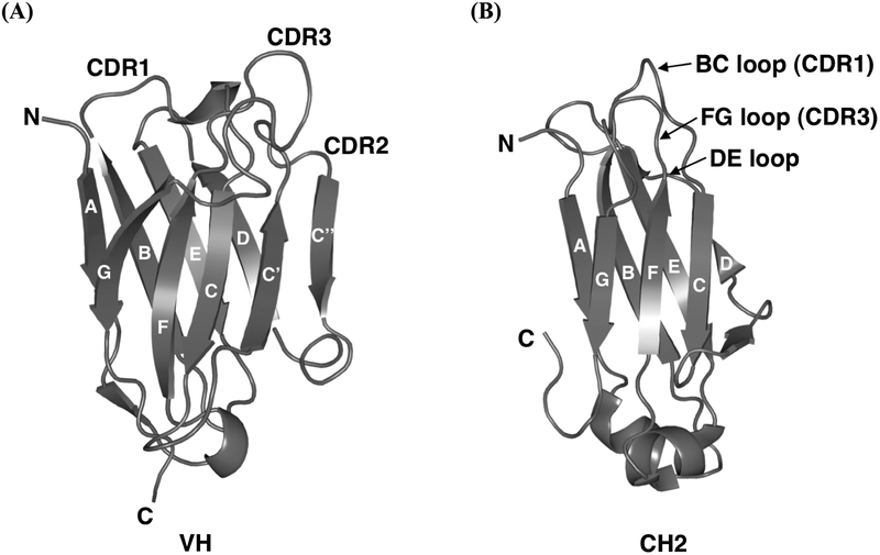
Fig. (2).
Ribbon diagrams illustrating (A) a model of the human antibody VH domain, m0, and (B) the crystal structure of the human IgG1 CH2 domain, which have been used as scaffolds for eAd library construction. The β-strands are labeled through A-G, having the same topology and similar structures except there are two additional strands, C′ and C″, in the VH domain. The CDRs or loop regions between those β-strands are at the same end of the barrel. The CDR1–3 in VH and BC, DE, and FG loops in CH2 are marked.

