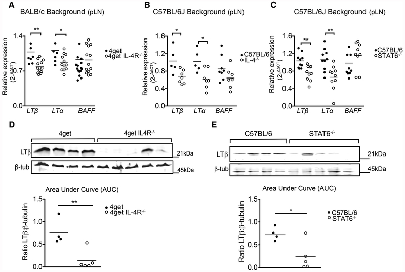
Figure 3.
LTβ and LTα mRNA levels in peripheral lymph nodes are dependent on the IL-4 signaling pathway in steady-state. 4get homozygous and 4get IL-4RαKO, as well as C57BL/6J and IL-4KO and STAT6KO mice were sacrificed at 6–9 weeks of age and popliteal lymph nodes were collected. Total RNA extracted using Trizol isolation followed by cDNA synthesis and quantitative expression of gene levels by q-RT-PCR. (A) Relative expression of LTβ, LTα, and BAFF normalized to beta-actin in 4get homozygous and 4get IL-4RKO mice. (B) Relative expression of LTα and LTβ and BAFF in whole popliteal lymph nodes from C57BL/6J and STAT6−/− and (C) C57BL/6J and IL-4KO mice. (D) LTβ protein expression normalized to β-tubulin in WT and 4get IL4RαKO mice. (E) LTβ protein expression normalized to β-tubulin in WT and STAT6KO mice. Statistical significance was determined by Man–Whitney test *p < 0.05, **p < 0.01. Data points represent individual mice. Quantitative PCR data represents three different experiments with n > 4 mice/group. Western blot data are representative of two independent experiments with n ≥ 4 mice per group.

