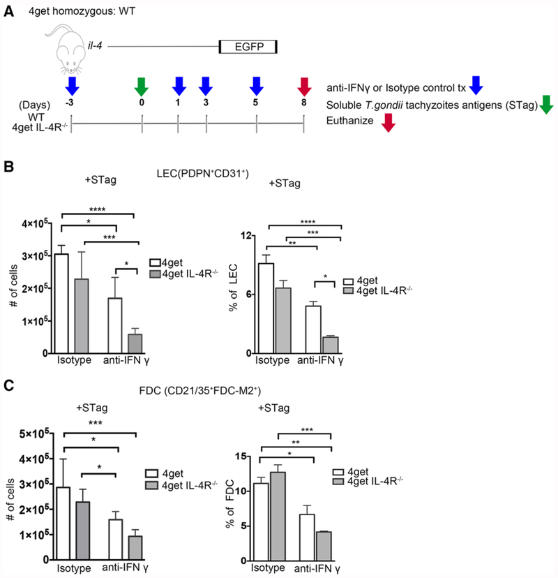
Figure 5.
IFN-γ promotes Stag induced LEC cell expansion in peripheral lymph nodes in the absence of IL-4Rα. 4get homozygous and 4get IL-4RαKO mice were immunized s.c. in the footpad with STag and treated with either anti-IFNγ or isotype control. Reactive popliteal lymph nodes were collected and single cell suspensions obtained and analyzed by flow cytometry. (A) Schematic of Stag immunization and blocking antibody treatment. (B) Cell numbers of lymphatic endothelial cells (PDPN+CD31+) in WT and knockout mice after treatment with isotype control or IFNγ blocking antibody, bars shown mean ± SEM. (C) Number of follicular dendritic cells (FDC-M2+CD21/35+) from isotype and IFNγ blocking antibody treated WT and knockout mice were analyzed by flow cytometry. Bars shown mean ± SEM, *p < 0.05, **p < 0.01, ***p < 0.001, ****p < 0.0001 (ANOVA, Bonferroni’s multiple comparison test). FACS data shown are concatenated from 3–6 mice per group, results are from two independent experiments.

