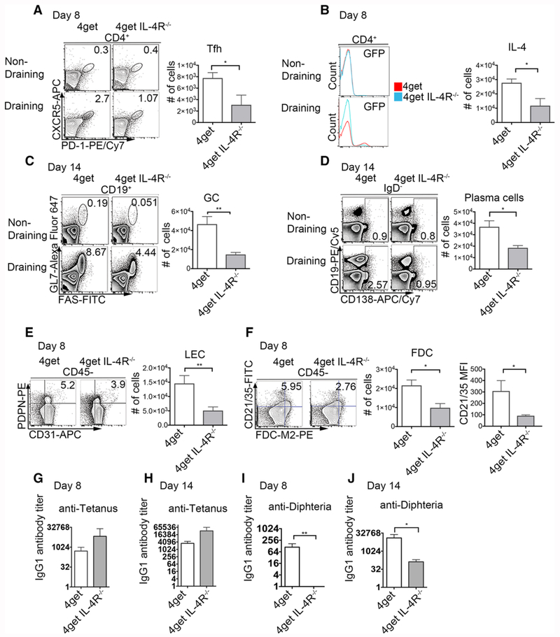
Figure 6.
Immunization with Tetanus/Diphtheria (TD) fails to induce Tfh expansion or a diphtheria-specific antibody response in mice lacking IL-4Rα. 4get homozygous and 4get IL-4RαKO mice were injected s.c. with 1/10 of the TD (with alum as adjuvant) dose employed in humans. Eight and 14 days postimmunization reactive peripheral lymph nodes were collected, digested, and filtered through a cell strainer to obtain a single cell suspension. Cells were stained with surface antibodies and analyzed by flow cytometry. (A) Expression and cell number of T-follicular helper cells (CD4+CXCR5+PD-1+) at day 8 from nondraining and reactive popliteal lymph nodes, bar denotes mean ± SEM (B) expression of IL-4 (GFP+) 8 days postimmunization with Tetanus diphtheria (C) Germinal center (CD19+GL7+FAS+) at day 14, (D) Plasma cells and plasmablasts (CD19+/− CD138+) at day 14 postimmunization pregated on IgD- (E) Lymphatic endothelial cells (PDPN+CD31+) at day 8 and (F) Follicular dendritic cells (FDCM2+CD21/35+) pregated on CD45 negative, cell numbers, and MFI, mean ± SEM. (G) Tetanus-specific IgG1 antibody titers at days 8 and (H) 14 postimmunization. (I) Diphtheria-specific IgG1 antibody titers at days 8 and (I) 14 postimmunization. FACS data represents concatenated samples from >3 mice per group and representative of three independent experiments for day 14 and two independent experiments for day 8. *p < 0.05, **p < 0.01; unpaired Student’s t-test.

