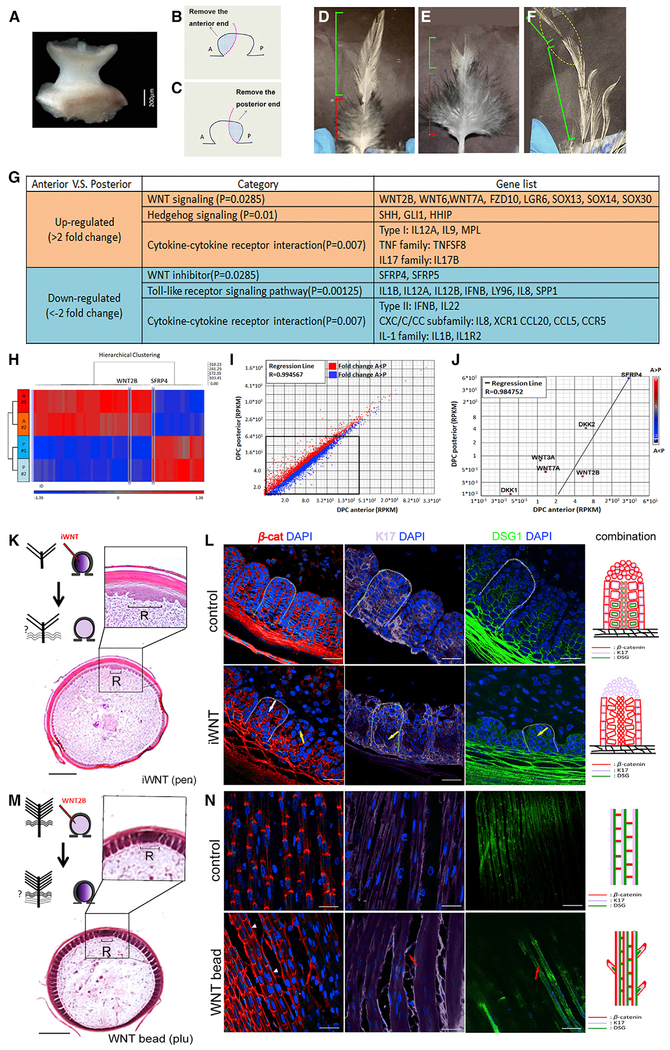
Figure 5. Asymmetric WNT Signaling from the Dermal Papilla Regulates Feather Branch Types by Altering Barbule Cell Shapes.
(A–F) Control of barbule morphology by DP niche. (A) Intact DP dissected from growth phase contour feather. (B and C) Schematic drawing shows the strategy used to ablate the DP anterior (B) or posterior (C) regions. Blue region is excised. (D) Normal contour feather with distal pennaceous and proximal plumulaceous barbs. Anterior DP ablation generated feathers that lost the distal pennaceous portion. (F) Posterior DP ablation produced feathers that lost the proximal plumulaceous portion. (D–F) Green bracket, pennaceous branch; red bracket, plumulaceous branch.
(G–J) Transcriptome differences of anterior versus posterior DP. (G) Signaling pathways up- or downregulated in the anterior compared with the posterior DPC region. (H) Heatmap of genes clustered in two groups, anterior versus posterior. (I) Scatterplots depicting transcriptomic comparisons between anterior and posterior DPC regions. (J) Differentially expressed WNT related molecules highlighted in scatterplots.
(K–N) Barbule cell organization alteration by WNT signaling perturbation. (K) Feather branch morphology after applying WNT inhibitor (iWNT) to anterior DP. (L) Barbs in iWNT-treated follicles show disorganized barbule cell arrangement and reduced (yellow arrow) or irregular (white arrow) molecular expression. (M) Feather branch morphology after WNT2B bead insertion to anterior DP. (N) Lateral longitudinal sections display extra hooklet-like structures (red arrow) and irregular β-cat expression pattern (white arrowhead) in plumulaceous region of treated follicles. Colors used in schematic drawings in (L) and (N) match those from Figures 4B and 4C. Scale bar, 200 μm (K and M) and 15 μm (L and N). A, anterior; β-cat; β-catenin; DAPI, 4′,6-diamidino-2-phenylindole; DPC, dermal papilla complex; DSG1, desmoglein 1; iWNT, Wnt inhibitor; K17, keratin 17; pen, pennaceous; plu, plumulaceous; R, rachidial zone; RPKM, reads per kilobase of transcript, per million mapped reads; P, posterior.
See also Figures S6 and S7.

