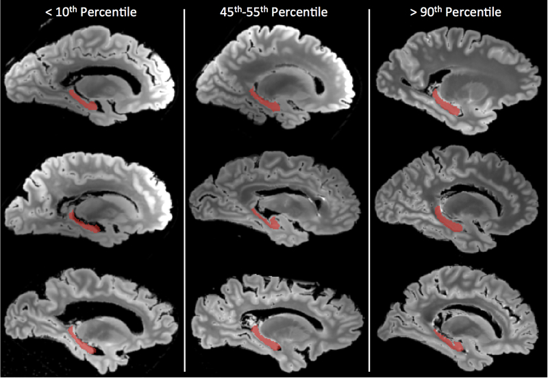
Figure 1.
Representative hippocampal segmentations (red) overlaid on sagittal slices of the shortest echo time image from a fast spin echo MRI pulse sequence carried out postmortem. Three ranges are represented: hippocampi with volume below the 10th percentile are shown in the left column, those between the 45th and 55th percentile in the middle column, and those above the 90th percentile in the right column. For each range, three cases were randomly selected for display.

