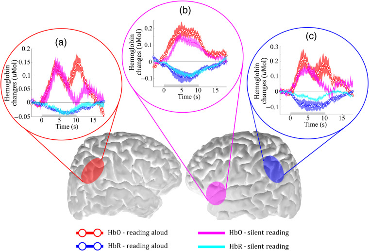
Fig. 5.
Temporal dynamics of the HRF due to reading tasks across all subjects from three different ROIs: (a) posterior region in the right hemisphere (contralateral to Wernicke’s), (b) Broca’s area, and (c) Wernicke’s area (left hemisphere) after applying our hybrid method to remove motion artifact. The gray bar on the plots indicates when the task was performed, and the shaded error bars are the standard error across all channels in the ROI. All ROIs are localized in the brain to facilitate visualization.

