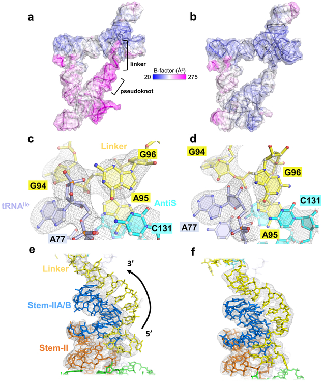
Figure 4.
Comparison of Mtb-ileS complex structures bound to tRNAIle-cP or tRNAIle-OH. a, Overall structure of Mtb-ileS bound to tRNAIle-cP with b-factor coloring. b, Overall structure of Mtb-ileS bound to tRNAIle-OH with b-factor coloring. c, Omit map electron density (gray mesh) at 3.0 σ of the aminoacylation sensing pocket in the tRNAIle-cP structure. d, Omit map electron density at 3.0 σ of the aminoacylation sensing pocket in the tRNAIle-OH structure. e, 2Fo-Fc map electron density (gray mesh) at 1.0 σ of the pseudoknot region in the tRNAIle-cP structure. f, 2Fo-Fc map electron density at 1.0 σ of the pseudoknot region in the tRNAIle-OH structure.

