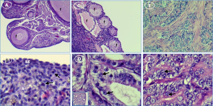
Fig 1. Hen ovary with and without tumors stained with H&E.
(A) normal ovary with developing follicles (f); (B) normal ovary showing surface epithelium (SE) and cells with resting nuclei (arrows) in stroma (s); (C) ovary with ovarian tumor has degenerating follicles (f); (D) activated nuclei (arrows) in mucinous tumor showing [inset, low magnification showing overall morphology]; (E) serous tumor; (F) higher magnification of E showing activated nuclei (arrows). Original magnification: (A) 4X; (B) 4X [inset, 40X]; (C) 10X; (D) 40X [inset, 4X]; (E) 10X; (F) 40XTumors commonly have a proliferative cell profile. Consistent with the ultrasound designation, several indicators of proliferation, PCNA (Proliferating Cell Nuclear Antigen) (p = 0.04), WT1 (Wilms Tumor 1 protein) (p = 0.012) and EpCAM mRNA (3.7x; p = 0.0001) were significantly higher in ovarian tumors compared to normal ovaries in the hen.

