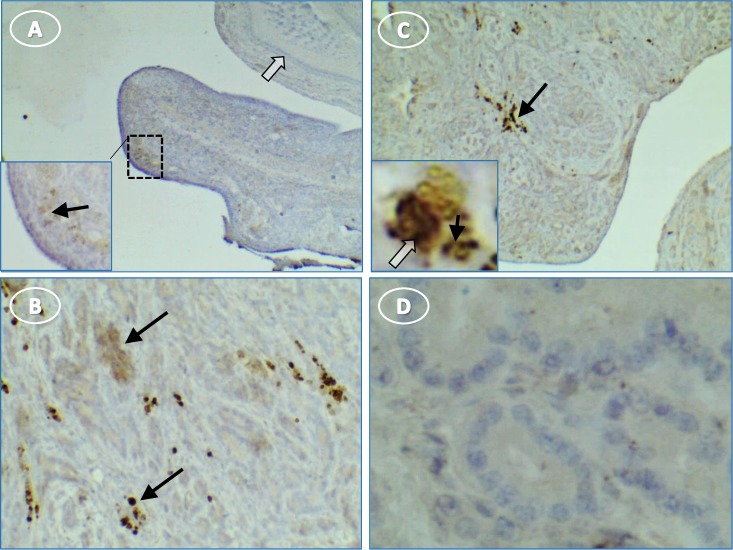
Fig 8. Hen ovary with and without tumors stained for IL1β.
(A) normal ovary without stain except some faint groups of cells [inset, higher magnification of A showing faint cells (arrow) near surface epithelium]; (B) stained groups (arrows) of immune cells in tumor ovary; (C) immune cell groups in tumor stroma and higher magnification inset showing punctate stain in cell cytoplasm (black arrow) and nuclear stain (white arrow); (D) control section of tumor without primary antibody is not stained. Original magnification: (A) 4X [inset, 10X]; (B) 10X; (C) 4X [inset, 40X]; (D) 40X.

