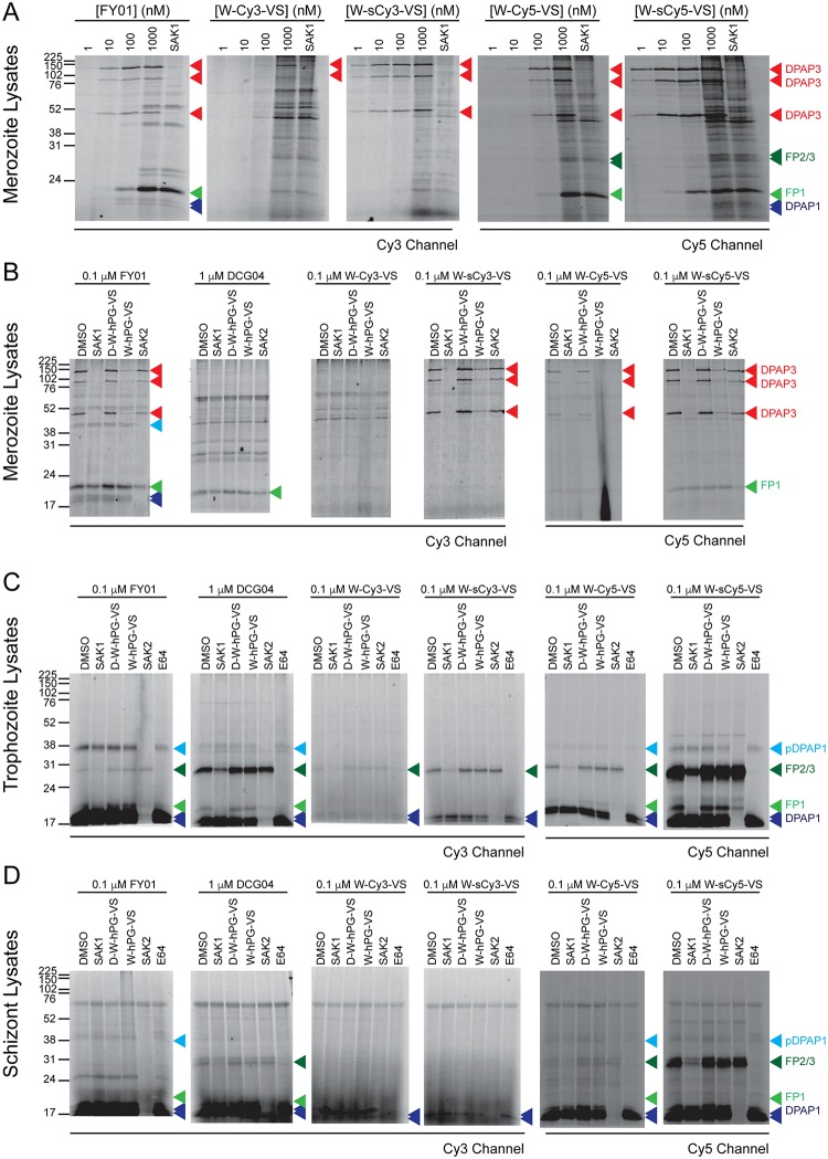
Fig 2. Labelling of cysteine proteases in parasite lysates.
(A) Merozoite lysates diluted 1:10 in acetate buffer were treated for 1 h with 1–1000 nM of the indicated ABPs. For the highest ABP concentration, samples were also pre-treated for 30 min with 1 μM of the DPAP3 inhibitor SAK1, which results in the loss of labelling of the three isoforms of DPAP3 running at 120, 95, and 42 kDa. (B-D) Lysates collected at merozoite (B), trophozoite (C), or schizont (D) stages were diluted in acetate buffer (pH 5.5), pre-treated for 30 min with DMSO or 10 μM of different known covalent inhibitors of DPAP1 (SAK2), DPAP3 (SAK1 or W-hPG-VS), the FPs (E64), or the negative control compound D-W-hPG-VS. This was followed by 1 h labelling with the different ABPs at 0.1 μM except for DCG04 that was used at 1 μM concentration. (A-D) The fluorescent bands corresponding to DPAP1, DPAP3, FP1, and FP2/3 are indicated by blue, red, light green, and dark green arrowheads, respectively. Two additional biological replicates of these experiments are shown in S3 Fig.

