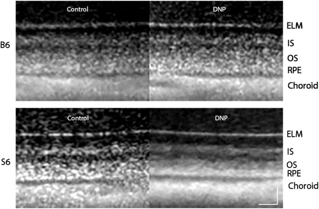
Fig 1. Representative same-eye OCT images focused on the superior outer retina layers for dark-adapted B6 and S6 mice before (Control) and after DNP treatment.
Layer assignments are: ELM, external limiting membrane; IS, rod inner segment layer; OS, rod outer segment layer; RPE, retinal pigment epithelium, as reported earlier [37]. Scale bars, 20 μm.

