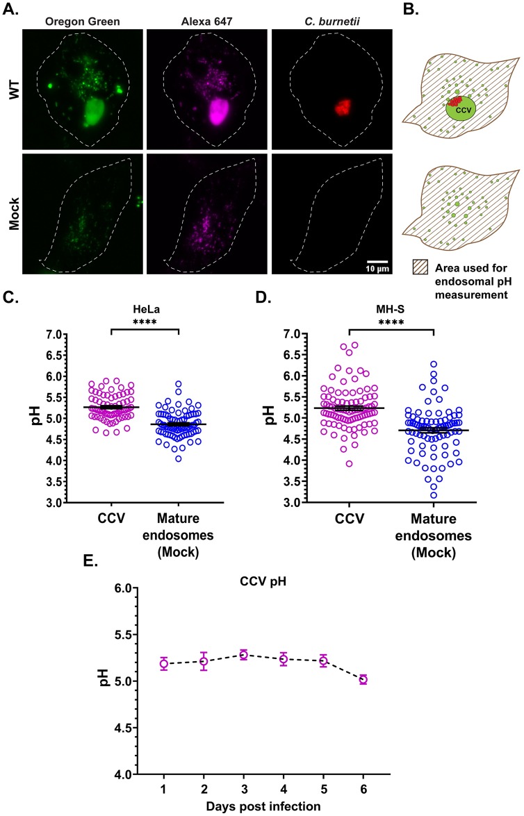
Fig 1. C. burnetii regulates CCV pH.
(A) Representative images of HeLa cells infected with mCherry-C. burnetii and pulsed with Oregon Green 488 (pH-sensitive; green) and Alexa 647 (pH-stable; magenta) dextran for 4 h followed by a 1 h chase. Z-stacks were acquired by live cell spinning disk microscopy and Oregon Green 488 and Alexa fluor 647 intensities quantitated. Oregon Green 488, which is quenched and less fluorescent at acidic pH, is visibly brighter in the CCV compared to the mature endosomes of mock-infected cells. (B) Diagram showing the area (patterned) used for endosomal pH measurement with the CCV excluded in infected cells. The endosomal pH is expressed as the average pH of all endosomes in the patterned area. (C, D) Ratiometric pH measurement in HeLa and MH-S cells at 3 dpi revealed CCVs are significantly less acidic than the mature endosomes of mock-infected cells. Data shown as mean±standard error of mean (SEM) of at least 20 CCVs or cells in each of three independent experiments as analyzed by unpaired student t-test; ****, P<0.0001. (E) Mean CCV pH from 1 through 6 dpi revealed that the CCV pH is relatively stable at pH~5.2 without any significant difference between the days, as analyzed by one-way ANOVA with Tukey’s posthoc test. Data shown as mean±SEM of at least 20 CCVs in each of three independent experiments.

