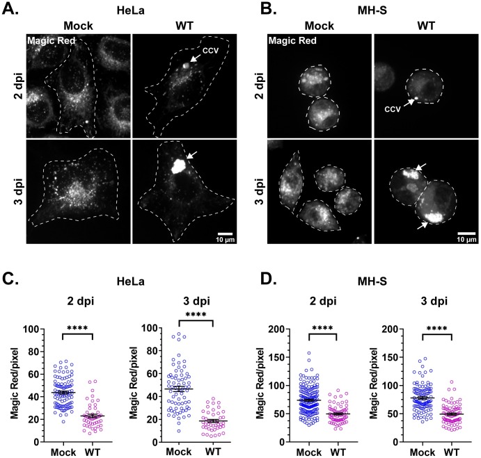
Fig 4. C. burnetii reduces proteolytically active lysosomes.
(A, B) Representative images of HeLa and MH-S cells stained with cathepsin B Magic Red to visualize proteolytically active lysosomes. mCherry C. burnetii-infected cells were plated in ibidi slides and labeled with Magic Red for 30 min followed by live cell confocal microscopy. (C, D) Quantitation of Magic Red intensity, normalized to cell area, revealed significantly less cathepsin B activity in WT C. burnetii-infected cells for both HeLa and MH-S cells at 2 and 3 dpi. Each circle represents an individual cell. Data shown as mean±SEM of at least 20 cells per condition in each of three independent experiments as analyzed by student t-test; ****, P<0.0001.

