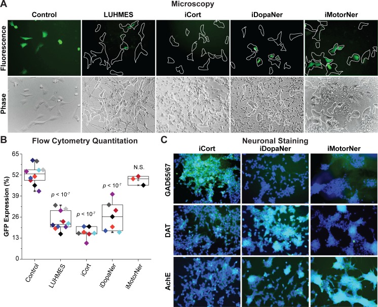
Fig 3. iPSC-derived neurons repress HIV expression.
(A) 60,000 hμglia/HIV HC69 cells were plated in the presence of 0.5 x 10⁶ LUHMES-derived neurons (as positive control) or 0.5 x 10⁶ iPSC-derived GABAergic cortical (iCort), dopaminergic (iDopaNer) or motor neurons (iMotorNer). HIV expression was evaluated after 24 h by fluorescence microscopy. Microglia identified by phase contrast microscopy are outlined by the white contours. (B) Flow cytometric analysis of microglial cell GFP expression. The p-values of pair-sample, Student’s t-tests comparing the microglial cells cultured alone or in the presence of neurons are shown. Individual independent experiments are color coded (n = number of independent samples). N.S.: non-significant. (C) Identification of differentiation of iPSC-derived neurons. Super-imposed images of DAPI stained nuclei (blue) and Alexa-Fluor 488 stained neuronal antigens (green) are shown. Neurons were stained with antibodies against GAD65/67, DAT, and AchE and Alexa Fluor 488-conjugated anti-rabbit secondary antibody.

