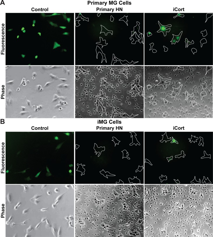
Fig 5. Primary neurons silence HIV expression in primary microglia.
(A) Human primary microglial (MG) cells. (B) iPSC-derived microglial (iMG cells). For each cell type, 60 x 103 cells were plated in the absence or presence of 0.5 x 10⁶ human primary neurons (Primary HN) or iPSC-derived GABAergic cortical neurons (iCort). HIV expression was evaluated after 24 h by fluorescence microscopy. Microglia identified by phase contrast microscopy are outlined by the white contours. Healthy neurons prevent spontaneous HIV reactivation in GFP- cells.

