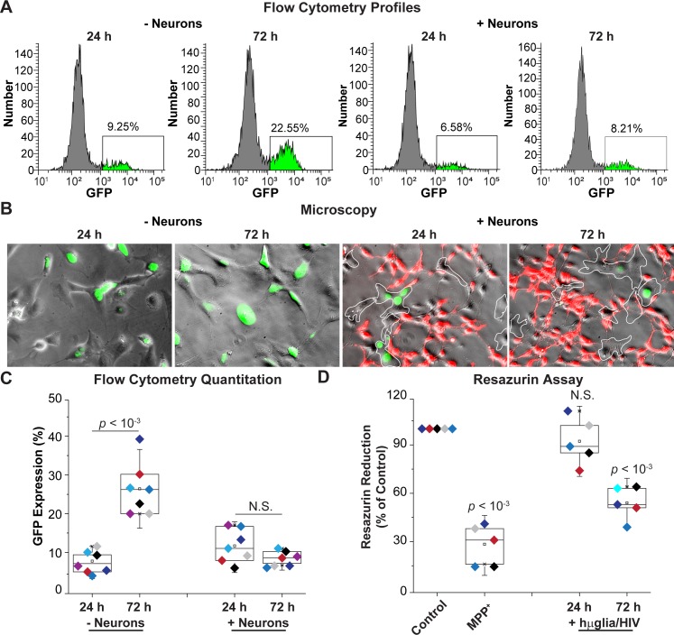
Fig 6. Neurons prevent HIV emergence from latency.
(A) Flow cytometry profiles of representative single cultures. hμglia/HIV HC69 cells were sorted into a GFP- cell population and cultured in the presence or absence of neurons. GFP expression was measured after 24 h or 72 h. (B) Microscopy of HC69 cells unexposed or exposed to neurons for 24 or 72 h. Microglial cells are outlined by a white dashed-line. (C) Quantitation of GFP expression. (D) Resazurin assay to evaluate neuronal viability. The resazurin reduction values (Y-axis) plotted are referenced to the control culture (neurons only), set at 100%. MPP+ was used a positive control for resazurin reduction. For both (C) and (D), the p-values of pair-sample t-tests of multiple experiments (n = number of independent samples) comparing the unexposed vs. the exposed cells are shown. N.S.: non-significant. Individual experimental series are color-coded.

