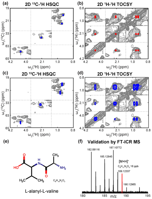
Figure 3.
Identification of L-alanyl-L-valine metabolite in mouse bile extracts. Panels a and b: 2D 13C-1H HSQC and 2D 1H-1H TOCSY of the unknown spin system A. Panels c and d: overlay of 2D NMR spectra of L-alanyl-L-valine (blue peaks) and bile extracts (gray peaks). Chemical shift agreement confirms the presence of L-alanyl-L-valine in the mouse bile mixture. Panel e: the chemical structure of L-alanyl-L-valine. Panel f: partial FT-ICR spectrum of the mouse bile extracts. The red peak with m/z 189.12337 of the [M+H]+ adduct is consistent with molecular formula C8H17N2O3 of L-alanyl-L-valine (mass error: 16 ppb).

