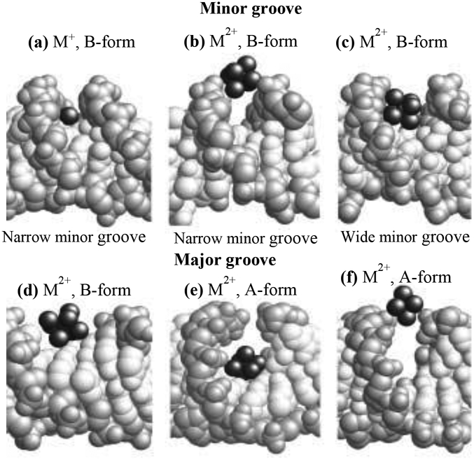
Fig. 1.
Space-filling models of X-ray crystal structures with cations bound in the minor and major grooves illustrating the correlation between DNA groove width and cation coordination sites. DNA base atoms are white and backbone atoms are light gray; cations and water oxygen atoms are dark gray (Reprinted from Ref,38 Copyright © 2001, with permission from Elsevier).

