Figure 5.
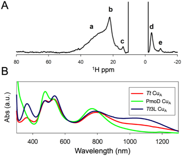
Paramagnetic NMR and electronic spectra suggest the absence of a thermally-accessible πu state in the PmoD CuA. (A) 600 MHz 1H NMR spectra of PmoD CuA recorded at 298 K in H2O. The broad signal a is observed after loss of the purple color, and is therefore attributed to a different Cu2+ binding site. Resonances b-e correspond to copper ligands of the PmoD CuA site. (B) Optical spectrum of PmoD CuA compared to those of subunit II of Tt CcO ba3 CuA and a loop mutant with a larger population of the πu state, Tt3L CuA.40

