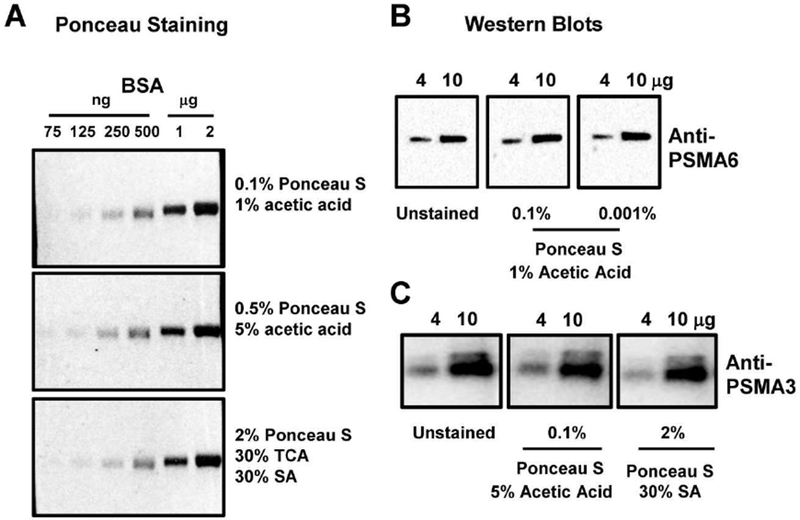
Figure 9.

Effect of Ponceau S stain on BSA detection and Western blotting Sensitivity. A) Quantification of 4μg and 10μg of protein stained with 0.1% (w/v) Ponceau S with 1% (v/v) acetic acid, 2% (w/v) Ponceau S with 30% (v/v) sulfosalicylic acid (SA), and 2% (w/v) Ponceau S with 30% (v/v) SA and 30% (v/v) trichloroacetic acid (TCA) for 2 minutes. B) Western blots of membranes stained with Ponceau S (0.01% and 0.001% in 1% acetic acid) and membranes not stained with Ponceau S. Western blots were carried out as described in the methods using anti-PSMA6 antibody. C) Western blots of membranes stained with 0.01% Ponceau S in 5% acetic acid, 2% Ponceau S in 30% SA and membranes not stained with Ponceau S. Western blots were carried out as described in the methods using anti-PSMA3 antibody.
