. 2019 Dec 19;8:e50279. doi: 10.7554/eLife.50279

Figure 4. The common activation mechanism is the shared portion of various downstream pathways of different class A GPCRs.

(a) Intracellular binding partners used in the active state structures. (b) Comparison of RRCS for active (green) and inactive (orange) states of eight receptors with different intracellular binding partners, including four recently solved cryo-EM structures of Gi/o-bound receptors (5-HT1B receptor, rhodopsin, A1R and µOR) (Tsai et al., 2018; García-Nafría et al., 2018; Kang et al., 2018; Koehl et al., 2018; Draper-Joyce et al., 2018) whose resolutions were low (usually ≥3.8 Å for the GPCR part). Nevertheless, almost all conserved residue rearrangements in the pathway can be observed from them. Three of 34 residues pairs were shown here, see Figure 4—figure supplements 1 and 2 for the remaining 31 residue pairs.

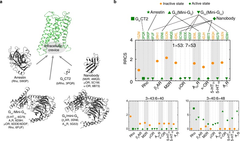
Figure 4.

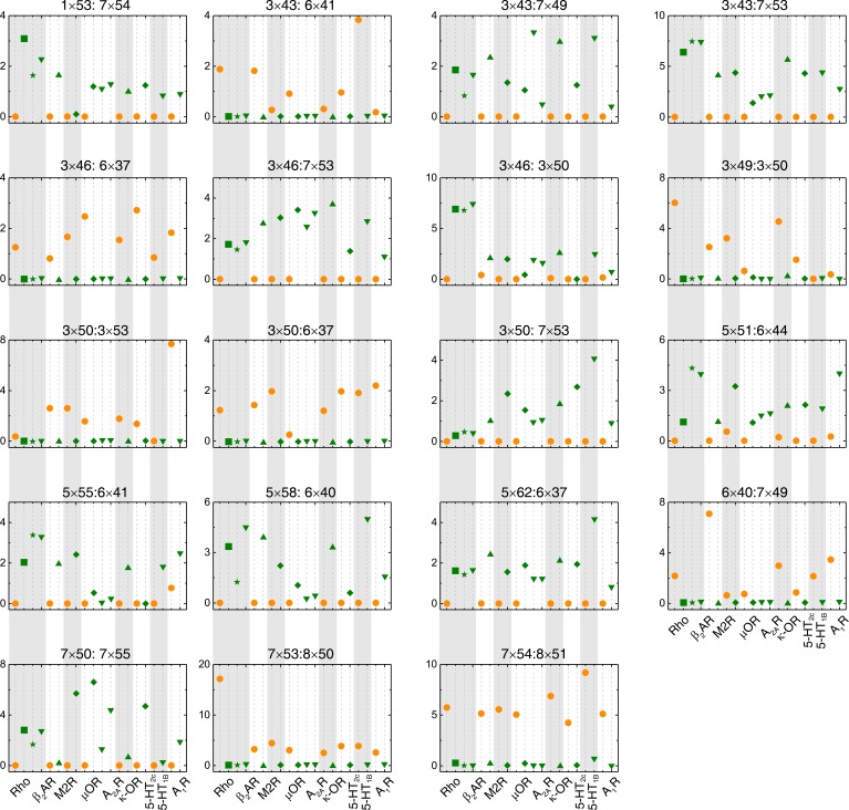
Figure 4—figure supplement 1. The switching conformation change is conserved upon receptor activation.

Figure 4—figure supplement 1.

Comparison of RRCS for active (green) and inactive (orange) states of eight receptors with different intracellular binding partners, including four recently solved cryo-EM structures of Gi/o-bound receptors (5-HT1B receptor, rhodopsin, A1R and µOR) whose resolutions were low (usually ≥3.8 Å for the GPCR part). Nevertheless, almost all conserved residue rearrangements in the pathway can be observed from them. Nineteen of 34 residues pairs were shown here, see Figure 4 and Figure 4—figure supplement 2 for the remaining residue pairs.
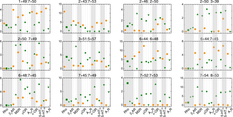
Figure 4—figure supplement 2. The repacking conformation change is conserved upon receptor activation.

Figure 4—figure supplement 2.

Comparison of RRCS for active (green) and inactive (orange) states of eight receptors with different intracellular binding partners, including four recently solved cryo-EM structures of Gi/o-bound receptors (5-HT1B receptor, rhodopsin, A1R and µOR) whose resolutions were low (usually ≥3.8 Å for the GPCR part). Nevertheless, almost all conserved residue rearrangements in the pathway can be observed from them. Twelve of 34 residues pairs were shown here, see Figure 4 and Figure 4—figure supplement 1 for the remaining residue pairs.