. 2019 Dec 19;8:e50279. doi: 10.7554/eLife.50279

Figure 5. Common activation model of class A GPCRs reveals major changes upon GPCR activation.

(a) Active and inactive state structures form compact clusters in the 2D inter-helical contact space: RRCSTM3-TM7 (X-axis) and RRCSTM3-TM6 (Y-axis). GPCR activation is best described by the outward movement of TM6 and inward movement of TM7, resulting in switch in the contacts of TM3 from TM6 to TM7. (b) Common activation model for class A GPCRs. Residues are shown in circles, conserved contact rearrangements of residue pairs upon activation are denoted by lines.

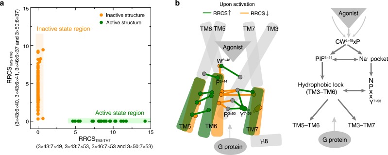
Figure 5.

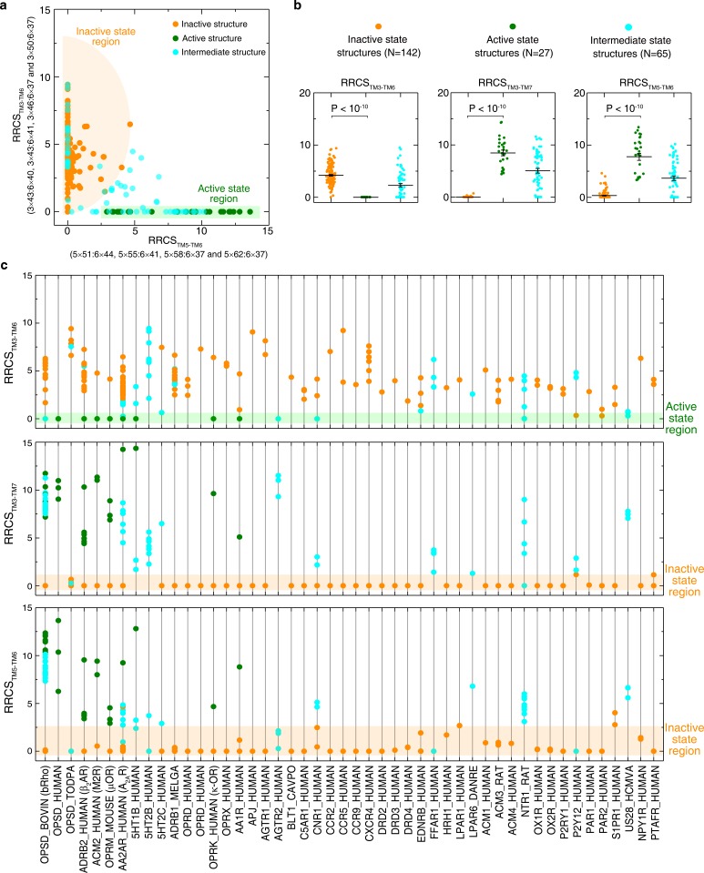
Figure 5—figure supplement 1. Global conformational change upon activation.

Figure 5—figure supplement 1.

(a) Distinct clustering of inactive- and active-state structures in two-dimentional inter-helical contact space RRCSTM5-TM6 vs. RRCSTM3-TM6. (b) The inter-helical contacts comparison between inactive- and active-state structures. (c) Receptor-specific inter-helical contacts for all class A GPCR structures (inactive, intermediate and active states are colored in orange, cyan and green, respectively). These results demonstrate that receptor activation involves the elimination of TM3-TM6 contacts, formation of TM3-TM7 and TM5-TM6 contacts, reflecting the outward movement of the cytoplasmic end of TM6 away from TM3, the inward movement of TM7 towards TM3 and the repacking of TM5 and TM6.
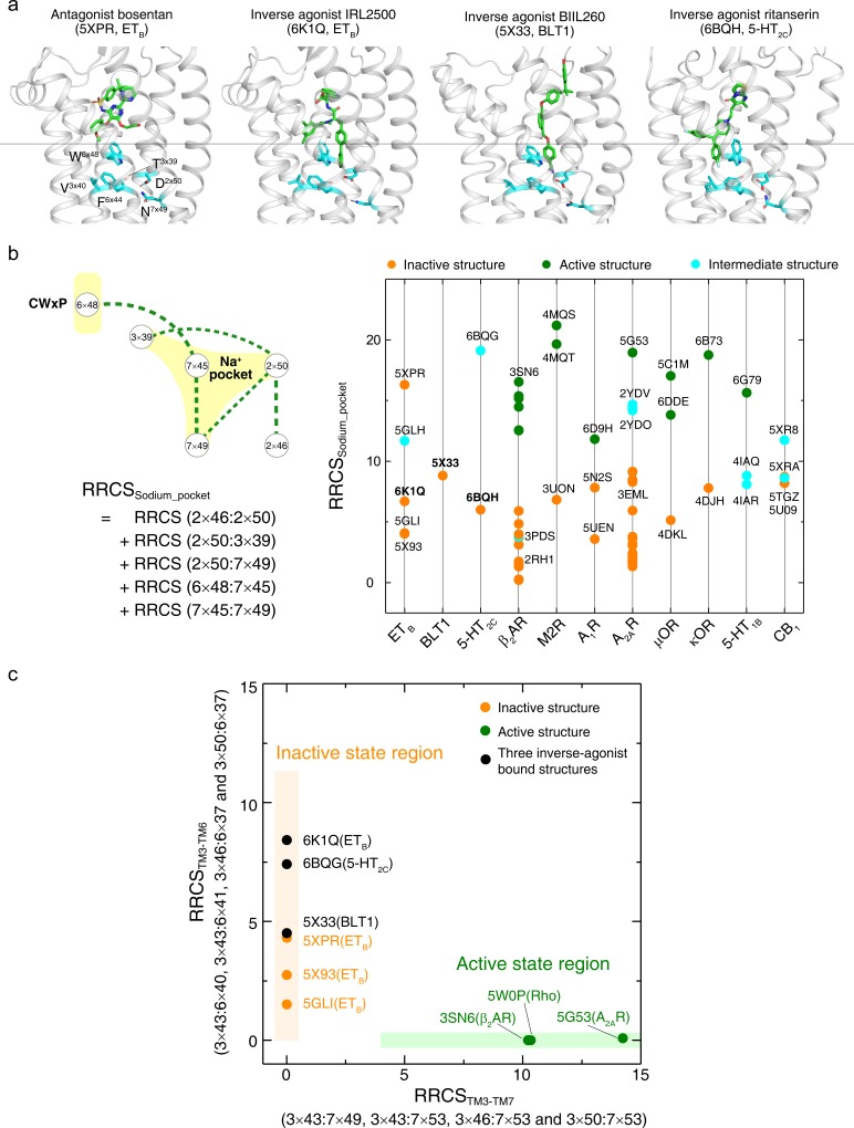
Figure 5—figure supplement 2. An inverse-agonism of class A GPCRs by preventing the collapse of Na+ pocket.

Figure 5—figure supplement 2.

(a) Comparison of binding poses of antagonist (bosentan [Shihoya et al., 2017]) and inverse agonists (IRL2500 [Nagiri et al., 2019], BIIL260 [Hori et al., 2018] and ritanserin [Peng et al., 2018]). Inverse agonists diffuse deeper in the ligand-binding pocket to touch the Na+ pocket. These key residues around the Na+ pocket are shown. (b) Comparison of the sum of contact scores of the conserved residue pairs around the Na+ pocket (RRCSsodium_pocket) between inactive- and active-state structures. The collapse of the Na+ pocket leads to a denser repacking of six residues (five residue pairs), reflected by higher RRCSsodium_pocket scores compared to that of the inactive state structures. (c) Distribution of three inverse agonist bound structures in the 2D inter-helical contact space: RRCSTM3-TM7 (X-axis) and RRCSTM3-TM6 (Y-axis). These inverse agonists bound structures (5×33, 6BQG and 6K1Q) located in the inactive state region with zero RRCSTM3-TM7 but high RRCSTM3-TM6 scores, despite deeper binding modes.