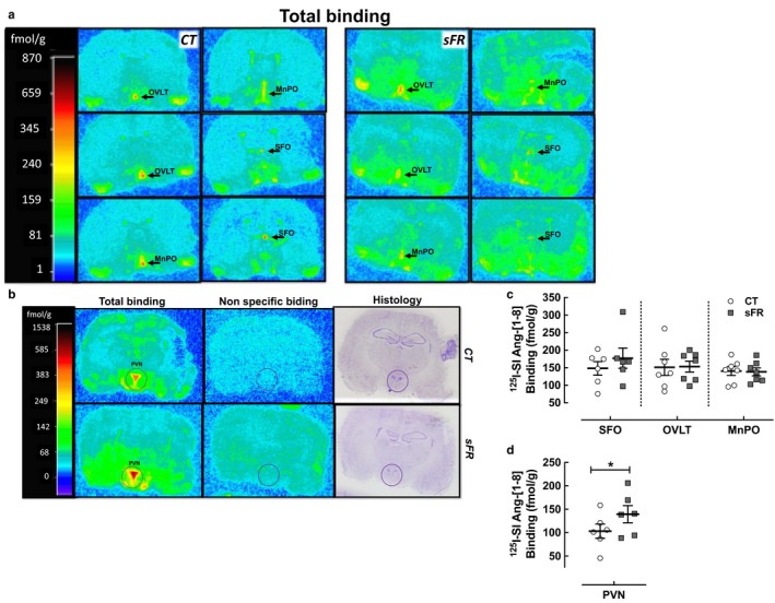
Figure 6.

Effect of severe food restricted (sFR) on AT1R binding in the AV3V and hypothalamus. Shown are representative images of total and nonspecific 125I‐[Sar1,Ile8]‐angiotensin‐[1‐8] (125I‐SI‐Ang‐[1‐8]) binding by in vitro autoradiography to brain slices containing the periventricular anteroventral third ventricle (AV3V) (a) and hypothalamus (b) on a CT or sFR diet. (a) The first top left panels are approximately ~0.3 mm rostral to Bregma, while the last right bottom panels are ~0.4 mm caudal to Bregma. Midline regions expressing highest AT1R binding (red color) are the organum vasculosum of the lamina terminals (OVLT), median preoptic nucleus (MnPO), and the subfornical organ (SFO) moving from rostral to caudal sections. Other regions displaying high AT1 receptor binding include the piriform cortex, ventral medial preoptic nucleus, lateral preoptic nucleus, and the suprachiasmatic nucleus. (c) Quantitation of specific 125I‐[SI]‐Ang‐[1‐8] binding in CT (open circle) and sFR (close square) rats (n = 7/group) is shown in the periventricular anteroventral third ventricle (AV3V) (d) and in the PVN. *p < .05 versus CT, by Student's t test. Values are expressed as the mean ± SEM
