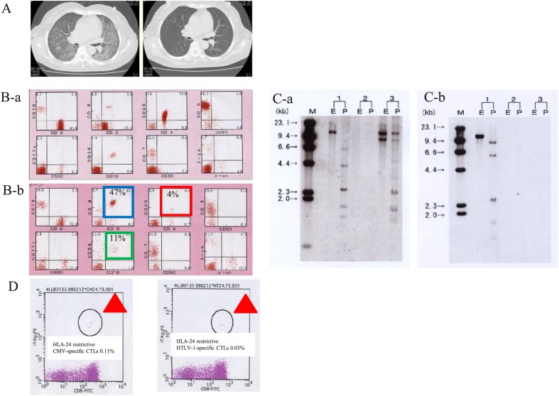
Fig. 5.
Clinical presentation of a spontaneous-remission case before and after treatment according to FCM analysis, Southern blotting, and CTL analysis.
Presented are the CT findings, FCM analysis, southern blot analysis, and CTL analysis by a tetramer assay in a representative case of spontaneous remission of ATL (case 6) after treatment of opportunistic infection (A-D). CT findings after treatment of CMV pneumonia showed the improvement of CMV pneumonia (A-a and A-b). FCM findings after treatment of opportunistic infection demonstrated the improvement of CMV pneumonia (B-a and B-b). Southern blot analysis after treatment of opportunistic infection showed the absence of HTLV-1 clonality in PB (C-a and C-b). Thus, CR was achieved because of the disappearance of ATL cells on smears of PB, FCM analysis, and Southern blot analysis after CMV pneumonia (A–D). Furthermore, case 6 had 47% CD8+ T cells and 11% NK cells according to FCM analysis of PB. In the HLA-A24 FCM tetramer assay, there were 0.03% HLA-24–restricted HTLV-1–specific CTLs and 0.1% HLA-24–restricted CMV-specific CTLs (D). Thus, CTL analysis by HLA 24 restrictive FCM analysis after treatment of opportunistic infection confirmed the presence of CMV-specific CTLs and HTLV-1-specific CTLs.

