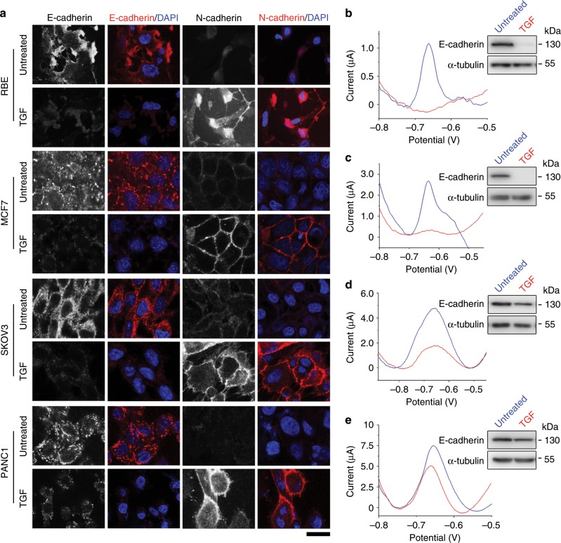
Fig. 7. The EMT biosensor is applicable in multiple cell lines.
a Immunofluorescence microscopy of E-cadherin and N-cadherin in RBE, MCF7, SKOV3, and PANC1 cells treated or untreated with TGF. Scale bar, 25 μm. b–e Electrochemical detection of RBE b, MCF7 c, SKOV3 d, and PANC1 e cells using the prepared biosensor. Upper right, immunoblots showing E-cadherin and α-tubulin levels in cells treated or untreated with TGF. Source data are provided as a Source Data file.

