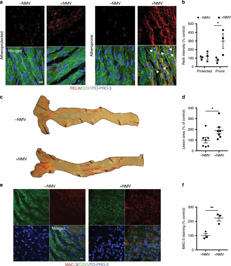
Fig. 8. NMVs induce activation of NF-κB in atheroprone regions and enhance atherosclerosis.
ApoE−/− mice fed a Western diet were injected with NMVs, or an equivalent volume of saline, twice weekly via the tail vein for 6 weeks. a Representative en face images of RELA (red) expression in the aorta of mice injected with saline (−NMV) or NMVs (+NMV) in atheroprone regions visualised using confocal fluorescence microscopy. Scale bar = 10 µm. Endothelial cells were identified by staining with anti-CD31 (green) and cell nuclei were identified using TO-PRO-3 Iodide (blue). Examples of nuclear staining of RelA indicated with arrowheads. b Mean fluorescence intensity was quantified using ImageJ software and data expressed as mean ± SEM (n = 5). c, d Plaque formation was measured in dissected aortae using en face Oil Red O staining and imaged by bright field microscopy. c Representative images are shown. d Areas of plaque formation were determined in the entire aorta using NIS-elements analysis software (n = 7). e, f The aortic arches of mice injected with saline (−NMV) or NMVs (+NMV) were studied by en face staining to quantify macrophages (MAC-3, red). e Endothelial cells were identified by staining with anti-CD31 antibody (green) and cell nuclei were identified using TO-PRO-3 Iodide (blue). Scale bar = 10 µm. f Mean fluorescence intensity was quantified using Image J software (n = 3). Data are expressed as a percentage of the mean of the control samples (−NMV) and presented as mean ± SEM. Statistical significance was evaluated using two-way ANOVA followed by Bonferroniʼs post hoc test (b) or an unpaired t-test (d, f). *P < 0.05, **P < 0.01. All n numbers represent independent animals. Source data are provided as a Source Data file.

