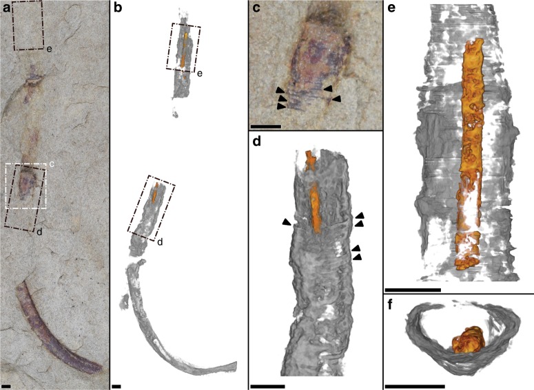
Fig. 4. Optical imaging and µCT of cloudinomorph pyritized tube and soft tissue.
a Light image of entire specimen (sample USNM-WCF_001) in plan-view, specimen partially obscured at rock surface. b Corresponding 3D volume render, showing soft tissue (orange) and tube wall (gray); boxes d, e are marked in both a, b to help guide slight differences in orientation. c Close-up view of labeled box in a, highlighting funnel rims (arrows) on external tube. d Close-up view of labeled boxes in a, b, 3D volume render showing partial soft tissue and funnel rims (arrows); d largely overlaps with c, but includes also host rock encased portion of the fossil. e Partial soft tissue from labeled boxes in a, b. f Cross-sectional view of e showing relative position of soft tissue that has settled to the bottom of the external tube wall. Sample reposited at the Smithsonian Institution. All scales = 2 mm.

