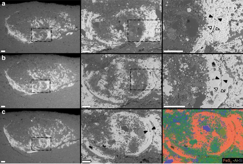
Fig. 6. Additional detail of cross-sectional morphology.
SEM backscattered electron micrographs (Z-contrast) of specimen USNM-WCF_001, as shown in Figs. 4, 5. Positioning of slices identified in Fig. 5f. Each row corresponds to a single slice at increasing magnifications from left to right (rows a–c); dashed boxes in left and middle columns correspond to location for higher magnification images. Right-most frame in row c shows EDS elemental map of middle frame in row c. Soft tissues in these slices are partially pyrite-infilled (increasingly so from a to c), though distinct sediment grains can be observed. Note also distinct soft-tissue wall boundaries, indicated by black arrows in higher magnification views. White arrows in higher magnification views of rows a, b indicate inferred direction of pyrite precipitation from soft-tissue wall, centripetally toward the interior and centrifugally from the exterior. Scales = 200 µm for left-most column, and 100 µm for middle and right-most columns.

