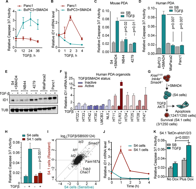
Figure 4: Dysregulated ID1 expression in PDAs with a functional TGF-β pathway.
A,B) Human PDA cell lines with wild type SMAD4 (Panc1) or with restored SMAD4 (BxPC3+SMAD4) were treated with 100 pM TGF-β for the indicated times. Apoptosis was measured by CaspaseGlo 3/7 and ID1 mRNA levels by qRT-PCR.
C) SMAD4-restored KrasG12D;Cdkn2a−/−;Smad4−/− mouse PDA cells (S4), and two KrasG12D;Cdkn2a−/− mouse PDA cell lines with wild type SMAD4 (NB44 and 4279) were treated with 2.5 μM SB505124 or 100 pM TGF-β for 36 h. Apoptosis was determined using CaspaseGlo 3/7. Mean ±SD, n=2, two-tailed unpaired t-tests.
D) SMAD4-restored BxPC3 human PDA cells, and the SMAD4 wild type MiaPaca2 and Panc1 human PDA cell lines were treated with 2.5 μM SB505124 or 100 pM TGF-β for 36h and assayed using CaspaseGlo 3/7 and CellTiter-Glo. Mean±SD, n=2, two-tailed unpaired t-tests.
E) S4, NB44, 4279, MiaPaca2, and Panc1 cells were treated with 2.5 μM SB505124 or 100 pM TGF-β for 24 h and subjected to ID1 and tubulin immunoblotting analysis.
F) qRT-PCR analysis of ID1 mRNA levels in human PDA organoids treated with or without TGF-β for 2 h. Mean±range of 3 replicates.
G) SMAD4-restored KrasG12D;Cdkn2a−/−;Smad4−/− mouse PDA cells were treated with 100 pM TGF-β and 2.5 μM MK2206 AKT inhibitor for 3 weeks, and surviving cells were selected.
H) SMAD4-restored KrasG12D;Cdkn2a−/−;Smad4−/− mouse PDA cells (S4) and a resistant population selected as described in G (S4.1 cells) were treated with 2.5 μM SB or 100 pM TGF-β, in the presence of 2.5 μM MK2206, for 36 h. Apoptosis was measured using CaspaseGlo 3/7. n=6 per group, mean ±SD.
I) S4 and S4.1 cells were treated with 2.5 μM SB or 100 pM TGF-β, in the presence of 2.5 μM MK2206, for 1.5h and subjected to RNA-seq analysis. Two replicates per sample.
J) S4 and S4.1 cells were treated with 100 pM TGF-β for the indicated times and Id1 mRNA level was determined by qRT-PCR. Mean±range of 3 replicates, representative of 2 independent experiments.
K) S4.1 cells with Tet-On shRNAs targeting Id1–3 were treated with 2.5 μM SB or 100 pM TGF-β, in the presence of 2.5 μM MK2206, for 36 h. Apoptosis was measured using CaspaseGlo 3/7. n=5 per group, mean ±SD, two-tailed unpaired t-test.

