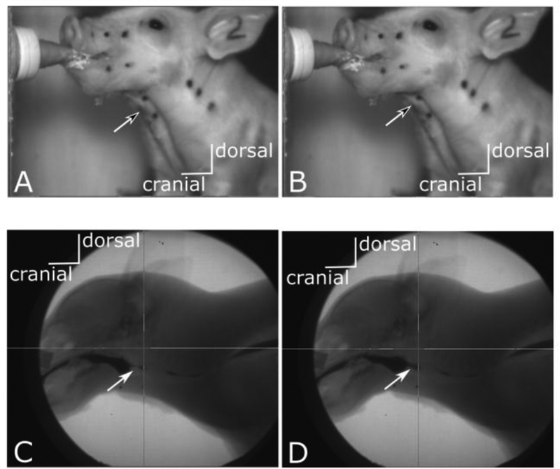
Figure 1:
Stills from external (top row) and videofluoroscopic (bottom row) videos of the same swallow in a pig. Left hand images (A, C) are from before swallow initiation. Right hand images (B, D) are those scored as marking the swallow. Black tipped arrows in the top row point to the laryngeal prominence and show elevation and change of angle with the hyoid in the left hand image. The two stills are two frames apart. White tipped arrows in the bottom row point to increase in milk volume in the pyriform recess in D, marking initiation of posterior bolus movement. Again, the two stills are two frames apart. Refer to supplemental materials for videos from which stills were taken.

