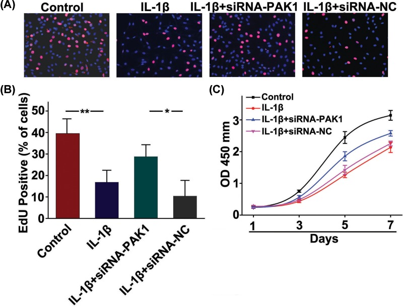
Figure 6. PAK1 is involved in IL-1β-inhibited rat chondrocytes proliferation.
(A) Representative photomicrographs of EdU staining. Red: EdU labeling of nuclei of proliferative cells (×200). (B) Quantitative data showing that the percentage of EdU-positive cells in different treatment groups (numbers of red vs numbers of blue nuclei); n = 6, *P < 0.05, **P < 0.01. (C) CCK-8 assay showed that cells viability was significantly inhibited in rat chondrocytes exposed to 10 ng/ml IL-1β from day 1 to day 7, whereas which was obviously alleviated by silencing of PAK1; n = 6.

