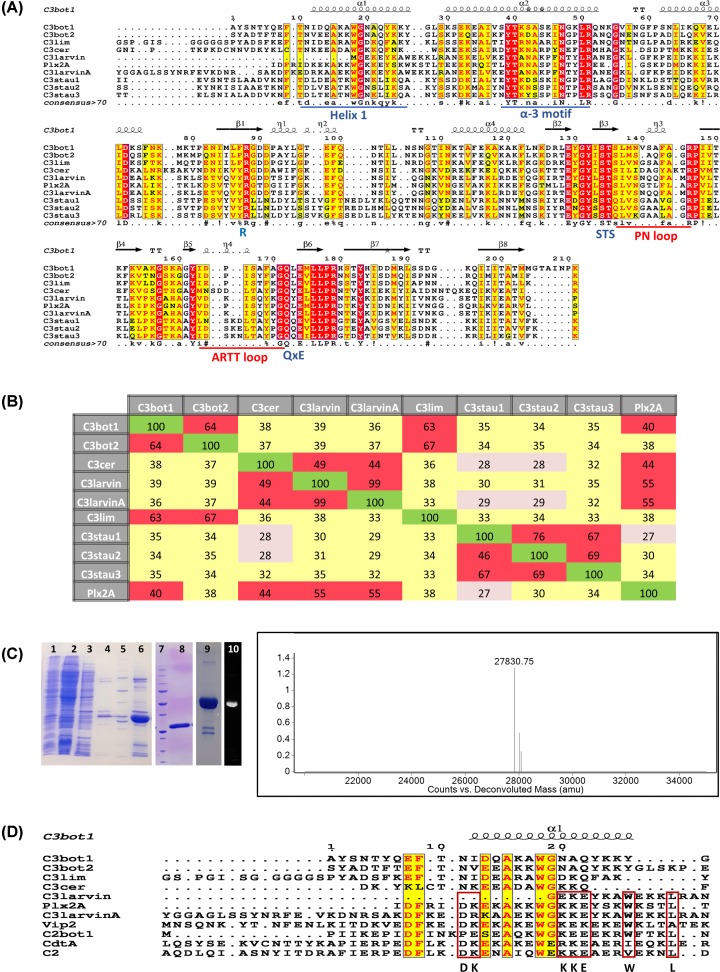
Figure 1. Multiple-sequence alignment of the C3 toxin subgroup.
(A) Multiple sequence alignment of C3 toxins and C3larvinA using the T-Coffee web server to align the sequences and ESPript to generate the figure [70]. Key catalytic regions are highlighted. Identical residues are highlighted in red, and similar residues are printed in red text and highlighted in yellow. The α-3 motif, helix 1, and the ARTT and PN-loops are indicated by underlined sequences. The three catalytic motifs in C3 toxins are indicated below the corresponding sequences. (B) Identity matrix showing the amino acid identity between the 100 core catalytic residues of the known C3 toxins and C3larvinA. Red, yellow and gray shading indicates high, medium and low sequence identities among C3 toxin pairs. The identity matrix was generated using ClustalX2 [71] and colored using Microsoft Excel. (C) Left side: purification and identification of C3larvinA from E. coli lysate. SDS/PAGE gels showing the purification and identification of C3larvinA. Lane 1, induced E. coli cell pellet; lane 2, IMAC sample flow-through; lane 3, column wash #1; lane 4, column wash #2; lane 5, Bio-Rad MW standards, 10-, 15-, 20-, 25-, 37-, 50-, 75-, 100-, 150-, 250-kDa; lane 6, partially purified C3larvinA as IMAC elution fraction; lane 7, Bio-Rad MW standards, Bio-Rad MW standards, 10-, 15-, 20-, 25-, 37-, 50-, 75-, 100-, 150-, 250-kDa; lane 8, purified C3larvinA after SEC; lane 9, Coomassie-stained RhoA reacted with C3larvinA and fluorescein-NAD+; lane 10, fluorescence image of lane 9 showing the fluorescence of the RhoA band. Right side: Q-TOF mass analysis of purified C3larvinA protein showing a single peak at 27830.75 Da, corresponding to the expected mass of recombinant C3larvinA (27830.66 Da). (D) Multiple sequence alignment of selected C2 and C3 toxins, and C3larvinA using the T-Coffee web server to align the sequences and ESPript to generate the figure [70]. Identical (or nearly so) residues between both C2 and C3 toxins are printed in red text and highlighted in yellow; identical (or nearly so) residues shared among the C2 toxins with P. larvae toxins, C3larvintrunc, Plx2A and C3larvinA, are bound by red rectangles. Abbreviations: IMAC, immobilized-metal-affinity chromatography.

