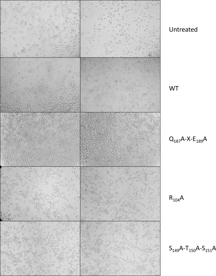
Figure 4. The effect of C3larvinA on cultured macrophages.
Cell morphology assays were performed with J774A.1 mouse macrophage cells that were grown to confluence in 25-cm2 culture flasks, the cells resuspended, and 100 μl was transferred to 6- or 96-well culture plates containing 4 ml supplemented medium (200 μl in the case of the 96-well plates). The cells were left for 48 h to grow in the new medium, at which point either toxin or control buffer was added. The cells were observed 20 h later and any morphology changes were recorded. (A) Untreated macrophage cells, (B) cells with buffer only; cells treated with: (C) 30 nM WT C3larvinA, (D) 300 nM WT C3larvinA, (E) 30 nM C3larvin A Q187A-X-E189A, (F) 300 nM C3larvinA Q187A-X-E189A, (G) 30 nM C3larvinA R104A, (H) 300 nM C3larvinA R104A, (I) 30 nM C3larvinA S149A-T150A-S151A and (J) 300 nM C3larvinA S149A-T150A-S151A. The elongated protrusions from the cells visible in (C) and (D) are phenotypic changes indicative of infection with C3 toxins. These protrusions are visibly absent for C3larvinA variant-treated cells (E–J).

