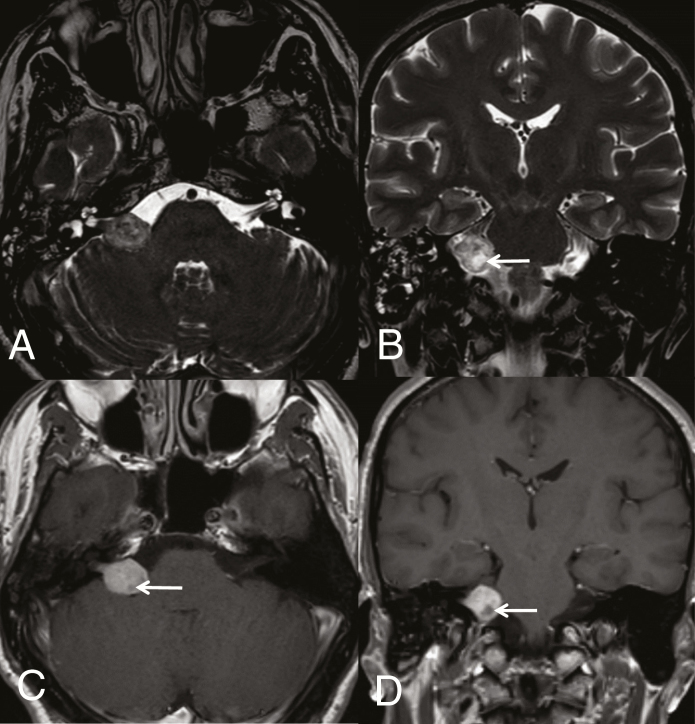
Fig. 1.
Intracanalicular and cisternal VS (Koos grade III). Axial 3D heavily T2-weighted sequence (DRIVE) (A) shows a VS expanding from the internal porus acusticus into the cerebellopontine-angle cistern. Coronal T2-weighted image (B) depicts slight mass effect on middle cerebellar peduncle. Cystic degenerative changes seen on T2 are well evident on axial (C) and coronal (D) T1-weighted images after gadolinium (arrows).

