Fig. 3.
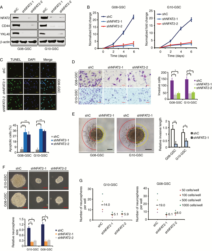
NFAT2 silencing inhibits MES GSC-enriched spheres clonogenicity in vitro. (A) Western blotting of NFAT2, CD44, and YKL40 in GSCs transfected with shRNA targeting NFAT2 (shNFAT2-1 or shNFAT2-2) or a control shRNA (shC). (B) Cell viability assay shows that NFAT2 knockdown markedly decreases the proliferation of G08 and G10 GSCs. (C) Targeting of NFAT2 via specific shRNAs significantly increases the apoptosis of G08 and G10 GSCs, as assessed with an assay by TUNEL (terminal deoxynucleotidyl transferase deoxyuridine triphosphate nick end labeling). Scale bar = 50 μm. (D) Representative microphotographs showing the invasion of G08 and G10 GSCs in the presence of NFAT2-shRNA or control-shRNA using the Matrigel assay. Cells were allowed to invade the Matrigel-coated filters toward the lower compartment for 20 h. Scale bar = 50 μm. Histogram showing the quantification of invasive cells. (E) The 3D spheroid invasion assay demonstrates that NFAT2 knockdown significantly decreased the invasion of G08 and G10 neurospheres. Scale bar: 100 μm. (F) Representative images of G10 and G08 neurospheres transfected with shRNA targeting NFAT2. Histogram showing the quantification of neurosphere size. Scale bar = 50 μm. (G) Limiting dilution neurosphere formation assays of effect of NFAT2 silencing on GSC renewal. Results are presented as mean ± SD of triplicate samples from 3 independent experiments. *P < 0.05 and **P < 0.01.

