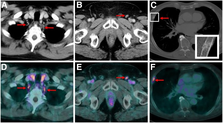
FIGURE 2.
CT images (top) and 18F-PSMA-1007 PET/CT images (bottom) of patient presenting with PSMA-ligand–positive, typically tear-drop–shaped cervical ganglia on both sides prevertebrally (A and D), unspecific PSMA-ligand uptake in nonenlarged left inguinal LN (B and E), and focal PSMA-ligand uptake in nondisplaced fracture of rib with corresponding fracture line and callus formation on CT images (C and F).

