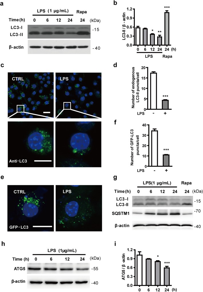
Fig. 1.
LPS inhibits autophagy in microglia. a The level of LC3-II, an autophagosome marker, declines in LPS-activated microglial cells. N9 microglial cells were stimulated with 1 μg/mL LPS for 6, 12, and 24 h. Cells were lysed and the levels of LC3-II (lipidated LC3) were analyzed by western blotting and quantified (b). c N9 microglial cells were treated with or without 1 μg/mL LPS for 24 h. PFA-fixed cells were stained with an antibody against LC3 and assessed by confocal microscopy to detect endogenous LC3-positive puncta (autophagosomes). Immunofluorescence images show the LC3-positive puncta (green) in CTRL- and LPS-treated cells. Nuclei are stained with DAPI (blue). Scale bar: upper: 20 μm, lower: 10 μm. d Statistical analysis of the number of endogenous LC3-positive puncta per cell in (c), from 3 independent experiments with at least 50 cells per treatment. e N9 microglial cells were transfected with GFP-LC3 plasmid for 24 h and treated with or without 1 μg/mL LPS for 24 h. GFP-LC3 puncta, representing autophagosomes, were observed by confocal microscopy. Representative images show GFP-LC3 (green) and DAPI (blue) in the CTRL and LPS-treated cells. Scale bar: 10 μm. f Statistical analysis of the number of GFP-LC3 positive puncta per cell in (e). g Primary microglial cells, isolated from rat brains were treated with LPS (1 μg/mL) for 6, 12, and 24 h or rapamycin (100 nM) for 24 h. Cells were lysed and the levels of LC3-II (lipidated LC3) and SQSTM1 were analyzed by western blotting. h N9 microglial cells were exposed to 1 μg/mL LPS for 6 h, 12 h, and 24 h. The expression of ATG5 was detected by western blotting and quantified (i). Data are presented as mean ± SEM. *p < 0.05, **p < 0.01, ***p < 0.001 vs control

