Figure 3.

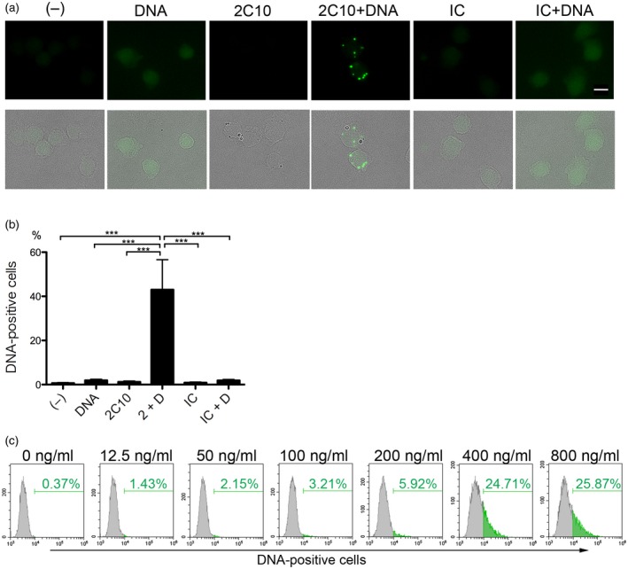
Anti‐DNA antibody 2C10 facilitates the internalization of DNA into THP‐1 cells. (a) THP‐1 cells were incubated with or without 400 ng/ml Alexa Fluor 488‐labeled DNA for 10 min, and then 10 μg/ml 2C10 or isotype‐matched control (IC) was added. After 2 h, internalized DNA was assessed. Upper row: DNA; lower row: merged DNA and transmitted light images. Scale bar = 10 μm. (b) Internalized DNA was quantified by flow cytometry and the mean ± standard error of the mean (s.e.m.) of the percentage of DNA‐positive cells was calculated from three independent experiments. ***P < 0·001, n = 5. (c) THP‐1 cells were incubated with the indicated concentrations of Alexa Fluor 488‐labeled DNA for 10 min, and then 10 μg/ml 2C10 was added. After incubation for 2 h, the percentage of DNA‐positive cells was assessed by flow cytometry. (–): no stimulation; 2 + D: 2C10 + DNA; IC + D: isotype control + DNA.
