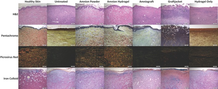
Figure 5.

Representative histological images for hematoxylin and eosin (H&E), pentachrome, picrosirius red, and iron colloid staining. H&E staining (row 1) demonstrated that amnion hydrogel and amnion powder had an epidermis very similar to that observed in healthy pig skin. Amnion hydrogel and amnion powder also had a very similar dermis organization compared with healthy skin. Untreated, hydrogel only, and AmnioGraft‐treated inconsistent and thinner epidermis, lacking noticeable epithelial rete peg protrusions. Hydrogel only‐treated wounds showed increased presence of darker‐stained larger fibers within the epidermis, but with minimal organization. Graftjacket‐treated wounds generally had an exposed dermis or were covered with a variably deep scab‐like tissue. Pentachrome staining (row 2) demonstrated that amnion hydrogel and amnion powder showed similar organized staining for collagen (yellow) and mature fibers (red) compared with healthy skin. Amnion hydrogel, amnion powder, AmnioGraft also showed mucins/GAGs staining (green). Untreated, hydrogel only, and Graftjacket‐treated tissues show primarily densely‐packed nuclei, yellow collagen staining, with some green staining for mucins and glycosaminoglycans. Graftjacket‐treated wounds were very cellular and showed little observable red, green, or yellow staining. AmnioGraft‐treated wounds demonstrated minimal red stained fibroid/muscle staining further indicating a lack of normal mature dermis structure. Picrorsirius red staining viewed with polarized light (row 3) confirmed observations from the pentachrome staining, demonstrating that amnion hydrogel and amnion powder‐treated wounds showed similar organized staining for mature collagen (red) and immature collagen (green) compared with healthy skin. Untreated and Graftjacket‐treated wounds did not stain positive for organized collagen structures. Hydrogel only‐treated wounds showed a moderate amount of positive staining for green and orange‐stained collagen, but significantly less than observed in healthy skin. Iron colloid staining (row 4) was performed to visualize Mucins/GAGs. Amnion hydrogel and amnion powder‐treated wounds showed intense iron colloid staining directly under the epidermis similar to healthy skin, whereas Graftjacket showed some intense staining under the inflamed scab area, as well as at the surface of the scab. Untreated, AmnioGraft, and hydrogel only‐treated tissues showed absent or minimal diffuse blue staining
