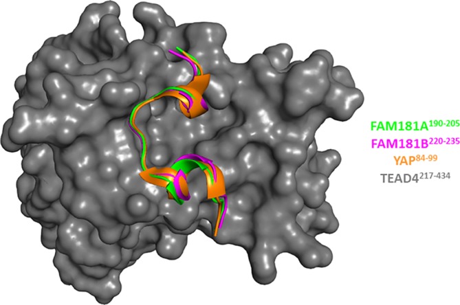
Figure 3.

Structures of the FAM181A190–205:TEAD4 and FAM181B220–235:TEAD4 complexes. The structures of the FAM181A190–205:TEAD4 (pdb http://bioinformatics.org/firstglance/fgij//fg.htm?mol=6SEN) and FAM181B220–235:TEAD4 complexes (pdb http://bioinformatics.org/firstglance/fgij//fg.htm?mol=6SEO) have been superimposed on that of the YAP60–100:TEAD4 complex (pdb http://bioinformatics.org/firstglance/fgij//fg.htm?mol=6GE3 26). FAM181A190–205, FAM181B220–235 and YAP84–100 are represented by green, magenta and orange ribbons, respectively. TEAD4 surface is colored gray. The picture was drawn with PyMOL (Schrödinger Inc., Cambridge, Massachusetts)
