Figure 3.

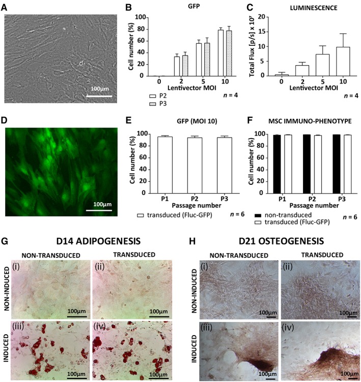
Characterization of non‐transduced and transduced rat ASCs. A, Brightfield image of non‐transduced ASCs in culture. B, GFP expression of transduced ASCs measured by flow cytometry with increasing MOIs as well as, C, luminescence measured by in vitro bioluminescence imaging. D, A fluorescent image of transduced ASCs (MOI 10) expressing GFP. E, GFP expression by ASCs transduced with an MOI of 10 in culture from passage 1 to 3 (P1 to P3). F, MSC immunophenotype (CD90+, CD29+, CD45−, CD31−) for transduced and non‐transduced ASCs. G, Qualitative assessment of adipogenesis for (i, ii) non‐induced control ASCs, (iii, iv) induced ASCs, (i, iii) non‐transduced ASCs, and (ii, iv) transduced ASCs all stained for oil red O to identify lipid droplet formation after 14 days in induction medium. H, Qualitative assessment of osteogenesis for (i, ii) non‐induced control ASCs, (iii, iv) induced ASCs, (i, iii) non‐transduced ASCs, and (ii, iv) transduced ASCs all stained for alizarin red S to confirm calcium deposition after 21 days in induction medium. Abbreviations: ASCs, adipose‐derived mesenchymal stromal cells; GFP, green fluorescent protein; MOI, mode of infection
