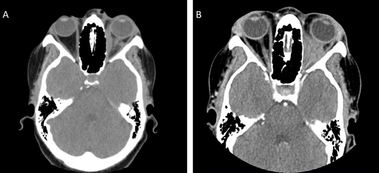
Figure 1.
(A. left): Previous CT imaging of the orbit and sella with intravenous contrast from a year ago indicating medial rectus oedema and inflammatory infiltration along with protrusion of the optic nerve. (B. right): Current CT imaging at the time of the first visit showing progression of retro-orbital inflammation and medical rectus oedema compared with previous year’s imaging.

