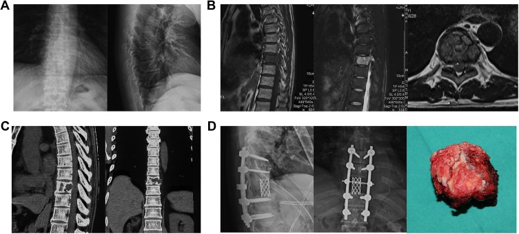
Figure 2.
A typical case underwent the removal of tumor by total en bloc spondylectomy in our center and was diagnosed as spinal metastasis from CCRCC. (A) Preoperative X-rays of anteroposterior and lateral spine demonstrated wedge deformation and osseous destruction in ninth thoracic spine. (B) Preoperative magnetic resonance imaging (MRI) indicated that the lesion showed low-intensity signal on T1-weighted image and high-intensity signal on T2-weighted image. (C) Preoperative CT showed osteolytic destruction in first lumbar vertebrae and its posterior elements, paravertebral soft tissue mass, and compression of spinal cord. (D) Total en bloc spondylectomy was conducted, and the ninth thoracic vertebral body was removed. The postoperative X-rays showed the ninth thoracic spine was removed and replaced by titanium mesh, with solid internal-fixation.
Abbreviation: CT, computed tomography.

