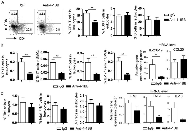
Figure 2. Activation of 4–1BB reduced the proportion of CD4 T cells and Th17 cells in the SMGs.
Anti-4–1BB antibody or IgG was i.p.-administered to 7-week-old female NOD mice 3 times weekly for 2 weeks, and the mice were analyzed 2 weeks after the last injection. (A) Dot plots, flow cytometry profile of lymphocyte populations; bar graphs, average percentage of lymphocyte populations in SMG leukocytes. (B) Left 4 panels, average percentage of Th17 and IL-17+ cells; right panels, relative gene expression of IL-23p19 and CCL20 levels in the SMG tissues, normalized to β-actin. (C) Left 3 panels, average percentage of Th1 cells, total IFNγ+ cells (including Th1, IFNγ + CD8 T/Tc1 cells, and all other IFNγ+ populations/subsets) and Tregs (Foxp3+CD4 T cells) among leukocytes; right panels, relative gene expression of IFNγ, TNFα and IL-10 levels in the SMGs, normalized to β-actin. Data are representative or the average of analyses of 8 mice per group. * p<0.05; **p<0.01.

