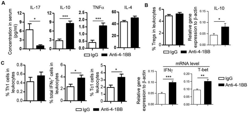
Figure 3. Activation of 4–1BB enhanced IFNγ production without affecting IL-17 production in SMG-draining lymph nodes and blood.
Anti-4–1BB antibody or IgG was i.p.-administered to 7-week-old female NOD mice 3 times weekly for 2 weeks. All the analyses were performed 2 weeks after the last injection. (A) Multiplex analysis of serum cytokine concentrations. (B) Left panel, average percentage of Tregs (Foxp3+CD4 T cells) in SGLNs as determined by flow cytometry; right panel, relative gene expression of IL-10 in SGLNs, normalized to β-actin. (C) Left 3 panels, average percentage of Th1, IFNγ+ and Tc1 cells in SGLNs; right panels, relative gene expression of IFNγ and T-bet in the SGLNs, normalized to β-actin. Data are the average of analyses of 8 mice per group. * p<0.05; **p<0.01; ***p<0.001.

