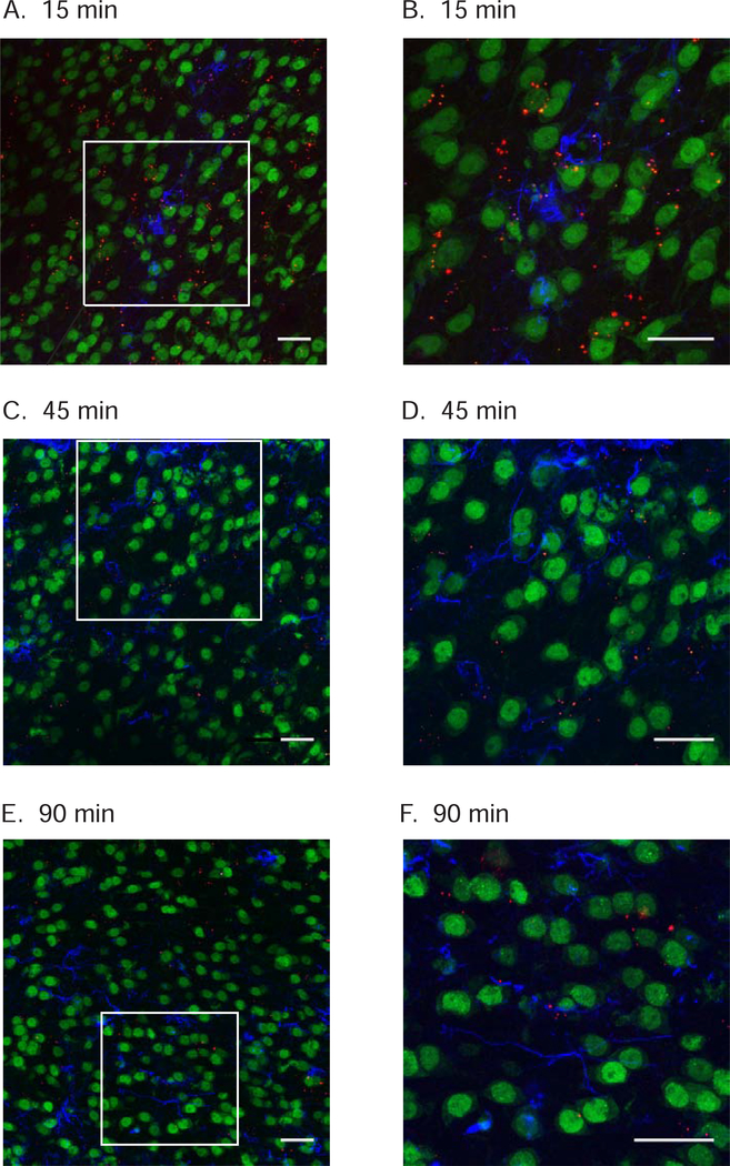
Figure 4.
TAMRA-conjugated TAT-P4-(DATC5)2 administered systemically crosses the blood brain barrier and is visualized in the nucleus accumbens shell. Separate cocaine-experienced rats were injected with TAMRA-conjugated TAT-P4-(DATC5)2 (3.0 nmol/g, i.v.) during extinction. Representative confocal images reveal TAMRA-conjugated TAT-P4-(DATC5)2 (red fluorescence) located in proximity to neurons labeled with NeuN (green fluorescence) and astrocytes labeled with GFAP (blue fluorescence) in the nucleus accumbens shell 15 (A), 30 (C) and 90 (E) minutes post infusion. Images shown in B, D & F are magnified inserts shown in A, C & E, respectively. Images are compressed z-stacks with a 0.5 μm step size (scale bar: 25 μm).

