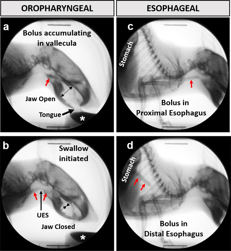
Fig. 1.
Videofluoroscopic swallow study (VFSS) assay. The oropharyngeal (a, b) and esophageal (c, d) stages of swallowing were assessed in the lateral plane while mice drank thin liquid contrast from a bowl (white asterisk). Jaw motion (open/closed cycles) and bolus flow (red arrows) were tracked frame-by-frame to quantitate several swallow metrics. UES upper esophageal sphincter

