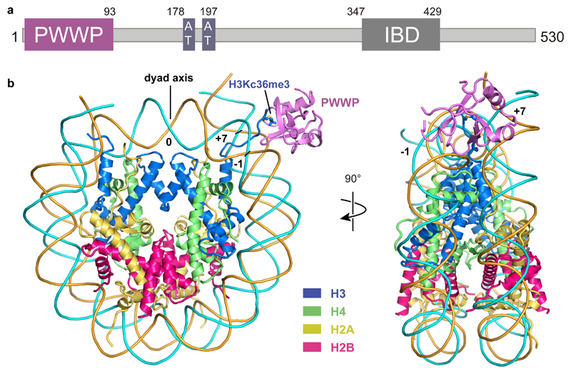
Figure 1. Structure of H3KC36me3-modified nucleosome bound to the PWWP domain of LEDGF.
(a) Domain architecture of LEDGF. Only the PWWP domain is visible in the structure. (b) Overview of the structure in front view (left) and side view (right). H2A, H2B, H3, H4, forward strand, reverse strand and LEDGF are colored in yellow, red, blue, green, cyan, orange and purple respectively. The color code is used throughout. H3KC36me3 residue is depicted as a stick model. SHLs 0, -1 and +7 are indicated with numbers.

