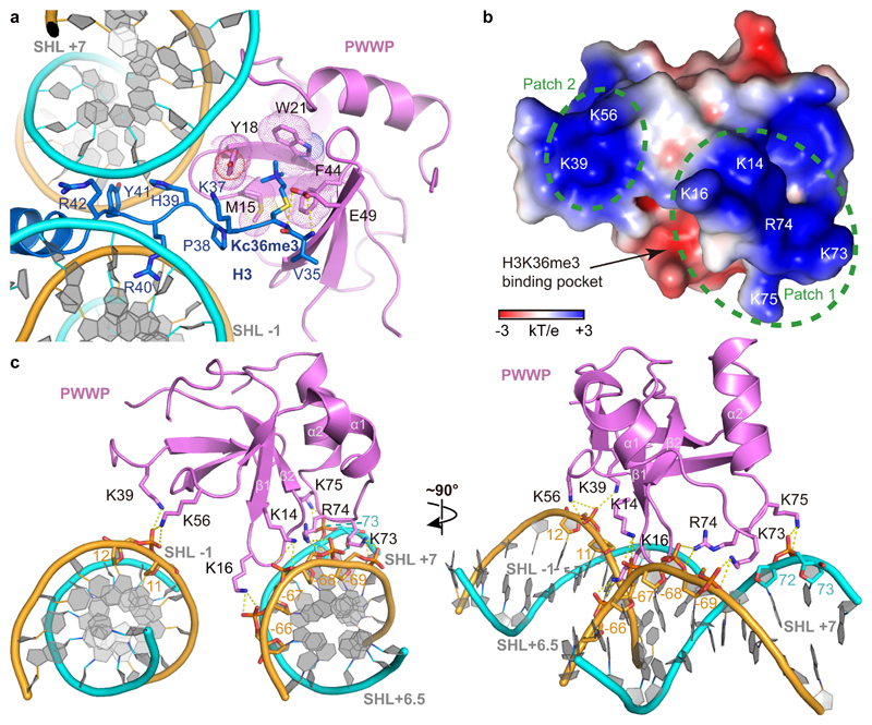
Figure 2. Nucleosome-PWWP interactions.
(a) Details of the interactions between PWWP and the methylated H3 tail. PWWP residues involved in H3KC36me3 recognition and H3 tail residues are shown as sticks. Selected hydrogen bonds are shown as yellow dashed lines. (b) Electrostatic surface of the PWWP domain calculated using the APBS tool in a range of -3 kT/e to +3 kT/e. Two positively charged surface patches involved in DNA interaction are indicated with green dashed circles; residues inside are denoted in white. (c) Details of DNA interactions. Electrostatic interactions and hydrogen bonds are shown as yellow dashes. SHLs are denoted. H3 tail is not shown for clarity.

